The National Food Imaging Centre: Equipment
Light Microscopy | Confocal Laser Scanning Microscopy | Electron Microscopy | Atomic Force Microscopy | Confocal Raman Microscopy | Image analysis and advanced software technologies | X-ray computed micro-tomography (micro-CT)
Light Microscopy
Conventional light microscopy remains a great, easy to use technique for analysing food structures. Information on particle size, shape, air content, crystal formation and ingredient distribution is quickly obtained using an appropriate contrast technique and interpretive skills. Our light microscopes are fitted with optical contrast techniques such as differential interference contrast to show phase separation behaviour in otherwise transparent liquids (Fig. 3), whilst dyes can identify specific components such as fats, proteins or polysaccharides. At the NFIC our conventional light microscopes can also be fitted with dynamic attachments such as temperature controlled stages and high speed cameras to allow for a vast range of experimental conditions and analysis.
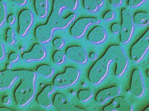
Figure 1: Light Microscopy: Protein/polysaccharide mixture showing two distinct phases
Confocal Laser Scanning Microscopy
Confocal Laser Scanning Microscopy (CLSM) is similar to epi-fluorescence microscopy, where a fluorescent dye is added to label the component of interest. General staining such as with protein and fat dyes can reveal ingredient distributions in real products. Various fluorescent staining techniques have been developed for a large range of food products and CLSM is a highly functional and valuable instrument pivotal to many projects and clients at the NFIC. A key feature of confocal microscopy is three-dimensional imaging of food structures. Additional features have been added to our microscopes including the ability to control heat and shear and a micro-tensile testing stage for solid foods. These types of attachments can facilitate the real-time study of food structures during conditions that might pertain to how food structures react during processing, cooking and mastication.
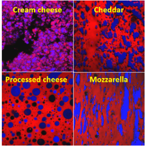
Figure 2: Cheese Analysis using Confocal Laser Scanning Microscopy (Red= Protein, Blue= Fat).
Electron Microscopy
An accelerated electron beam has a much smaller wavelength; consequently, much higher resolution is possible. The electron beam is focused by electromagnetic “lenses” and images are formed either by scanning the surface of a bulk sample such as in scanning electron microscopy (SEM); or by looking through a very thin sample as in transmitted electron microscopy (TEM). Traditionally, electron microscopy of food samples required extensive sample preparation involving chemical fixation, dehydration and possibly resin embedding. However, recent advances in the technology, in particular field emission electron optics, low-vacuum technology and cryo-preparation mean that it is now possible to visualize “difficult” food samples close to their natural state at a resolution of a few nanometers and with little or no sample preparation. These include liquids, frozen products and samples with high moisture, fat or sugar contents such as yoghurt (Fig. 6), dairy spreads and ice cream. The SEM at Moorepark uses field emission technology and is fitted with secondary, backscattered and transmitted electron detectors and most importantly the cryo-stage. An additional variable pressure mode allows examination of partially hydrated samples.
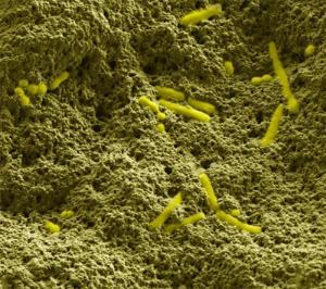
Figure 3: Cryo-scanning electron microscope image of yoghurt showing rod and cocci-shaped starter culture bacteria (yellow) embedded in a matrix of milk protein particles.
Atomic Force Microscopy
AFM is very different from the other imaging techniques described above, in that images are generated by moving a very small silicon nitride tip across the surface of the sample. The tip is attached to a cantilever, which deflects in response to minute surface variations in height. The deflection is converted to a brightness value that we see on the screen. This technique provides the highest possible imaging resolution of biological materials, permitting direct visualization of individual biomolecules including lipids, proteins and polysaccharides as well as microorganisms. A resolution of one nanometer has been achieved on biological samples such as DNA. A unique feature of AFM is its ability to characterize the mechanical properties of food materials, including stiffness, elasticity, friction and stickiness in addition to surface topography. Samples can be analysed under ambient conditions in liquid or gas environments and over a wide range of temperatures.
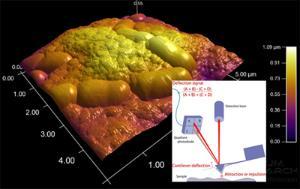
Figure 4: Atomic force microscope image of bacteria (Lactobacilli) embedded in whey protein gel. Inset shows the principle of the technique.
Confocal Raman Microscopy
Confocal Raman microscopy is a powerful technique to provide simultaneous microstructural and chemical information of food materials without the need of staining the samples. This technique is based on the optical principles of confocal microscopy, allowing optical sectioning of the samples and reconstruction of 3D stacks, and the physic-chemical principles of Raman spectroscopy, which allow identifying food ingredients based on their characteristic spectra.
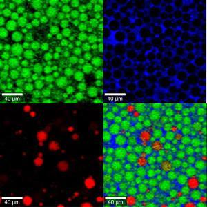
Figure 5: Confocal Raman micrograph of a mixed oil-in-water emulsion containing β-carotene in some of the oil droplets. The image shows the distribution of oil (green), β-carotene (red) and the aqueous phase (blue).
X-ray computed micro-tomography (micro-CT)
X-ray computed micro-tomography (micro-CT) is a non-destructive, three-dimensional imaging technique based on the differences in X-ray attenuation of the various components within a sample. It allows obtaining bi-dimensional cross-sections of materials and their 3D reconstruction without physically sectioning them. Compared to traditional CT scanners, micro-CT scanners can achieve sub-micrometric resolutions. In food, this technique can be used to study a wide range of samples, especially those that contain features with distinct densities. The obtained virtual 3D representation of the samples can be used to perform accurate measurements in any direction within the sample, using quantitative image analysis or even augmented reality tools. Our multiscale scanner has three different detectors for maximum flexibility in the type of materials, scales and sample sizes that can be analysed. The system also counts on in-situ temperature controlled stages to perform experiments at specific temperatures, and a mechanical loading stage to perform tensile/compression experiments and study mechanical-structural relationships in food.
Image analysis and advanced software technologies
Outputs from the various microscopes are all in the form of digital images and these can be analysed using a wide range of sophisticated image analysis software packages. Typical measurements would include particle size, phase volume, clustering or alignment. Image analysis data can then be correlated with other physical measurements and compositional data. Images are generated into full client reports which are then stored on a secure fully networked server-based image management system.
At the NFIC specialised softwares incude high speed video imaging softwares and volumetric 3D and 4D virtual reality software which are used to generate structural information about products once image sets are established with other techniques. High level of visualisation and data outputs are an exciting development as we merge conventional microscopy methods with advanced computerised techniques.